

Computed tomography (CT) scans, Magnetic Resonance Imaging (MRI) and X-rays are all diagnostic tools that allow doctors to see the internal structures of the body. These imaging tests are generally non-invasive, painless, and relatively quick.
However, diagnostic imaging tests are not the same. Each uses a different technology to create images and detect different types of medical problems. They also differ widely when it comes to accessibility, level of detail in the images and the type of energy used.
X-Rays
An X-ray, also called a radiograph, is a quick, painless procedure that typically takes a few minutes to complete. They are the most widely available and commonly used diagnostic imaging tests. Even when doctors want to order multiple tests, they will likely order an X-ray before more sophisticated tests such as CT scans and MRIs.
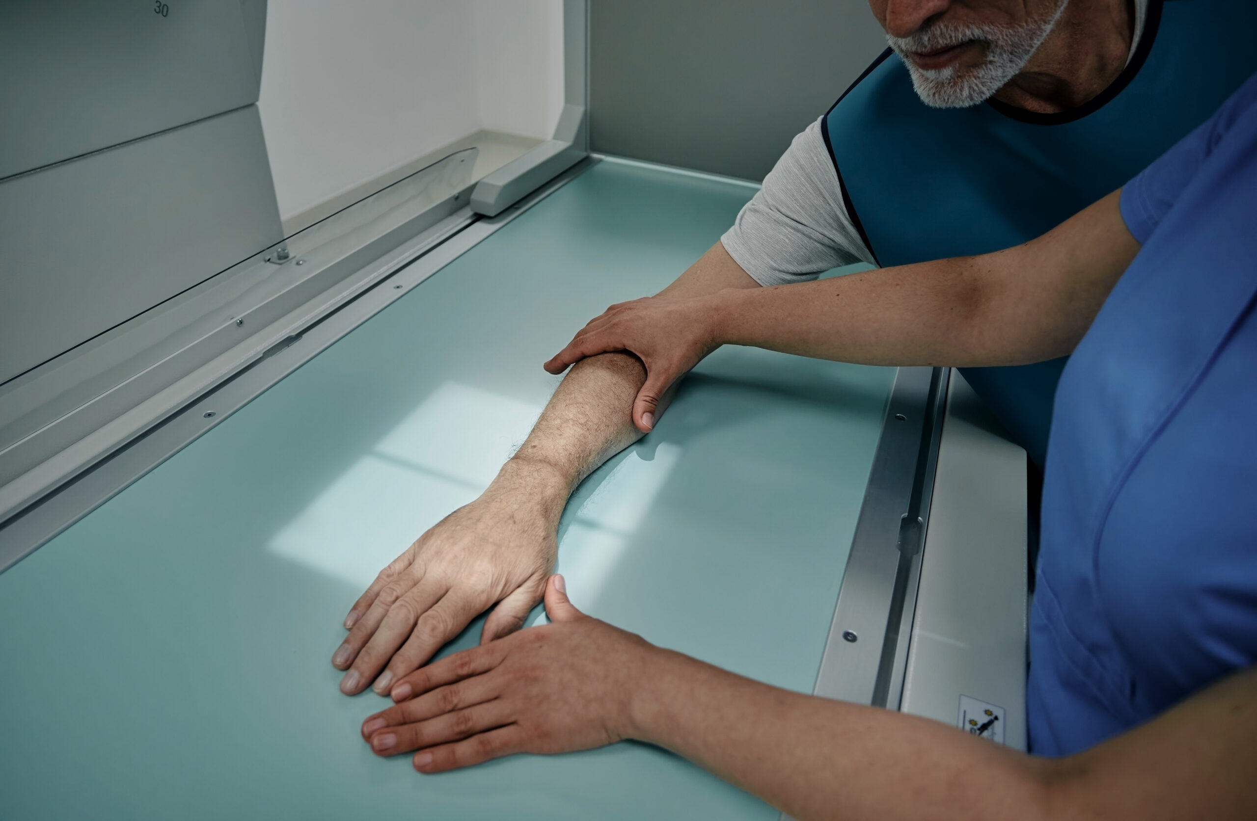
An X-ray sends radiation through the body. Areas with high levels of calcium, such as bones and teeth, block the radiation, causing them to appear white on the image. Soft tissues allow the radiation to pass through. They appear gray or black on the image. The level of radiation exposure from x-rays is not harmful, but your doctor will take special precautions if you are pregnant.
“An X-ray exam is usually the first-line imaging for acute injury or chronic condition,” says Keith Garner, Executive Director of West Tennessee Imaging Center. “X-rays often allow us to see major problems with the bones or rule out a fracture. However, they often will not show subtle bone injuries, soft tissue injuries or inflammation.” That is when a provider may order more advanced imaging for better detail of suspected injury or other concerns.
CT Scans
CT scans are kind of like the 3D version of an X-ray, but they cost more and take more time. A CT scan generates cross-sectional images. It offers a much higher level of detail, creating computerized, 360-degree views of the body’s structures. A CT scan allows the Radiologist (MD specializing in Imaging), to see the size, shape and position of structures that are deep inside your body. This includes:
- all bones
- organs
- tissues
- tumors
CT scans can spot blood clots, bone fractures, including subtle fractures not visible on X-ray as well as organ injuries. It can be done in either a hospital setting to aid in diagnosing illness or Emergency Department, for traumatic injury as well as an outpatient imaging center.
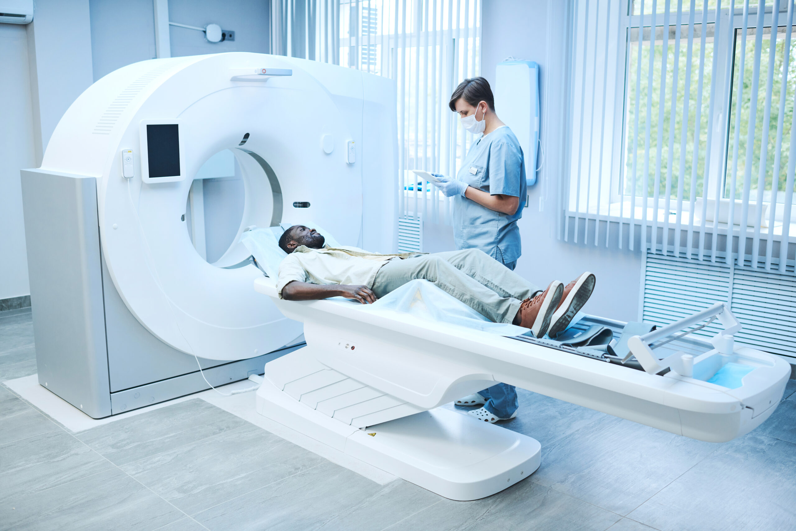
During a CT scan visit you may be asked to drink a fluid that helps the Radiologist see you GI tract and you may also have an IV started in your vein for an injection IV contrast that aids in viewing the organs within your body. You will lie on a flat, sliding mechanical table that is padded and when the scan starts, the table will pass into a ring-shaped CT scanner that will rotate and take a series of X-rays from different angles. You may hear a whirring sound from the CT machine. The scan will only take minutes to complete. A computer combines in real time, the images to produce a clear, two-dimensional view on a computer monitor. CT scans are painless and detailed other than a slight warm feeling within your body from the injection of the IV contrast. They take longer than X-rays but are still fast which makes them ideal for emergency situations.
CT scans and MRIs are both used to capture cross-sectional images within your body. The biggest difference is that MRIs use magnetic fields and CT scans use X-rays. While both are relatively low risk, there are differences that may make each one a better option depending on the circumstances. CT scans are more widely used than MRIs and are typically less expensive. MRIs, however, are thought to be superior regarding the detail of the image.
A CT scan may be recommended if a patient can’t have an MRI. People with metal implants, pacemakers or other implanted devices shouldn’t have an MRI due to the powerful magnet fields inside the machine unless the device is properly reviewed and ok’d to withstand and MRI exam. While CT scans create images of bones and soft tissues, they aren’t as effective as MRIs at exposing subtle differences between types of tissue.
MRIs
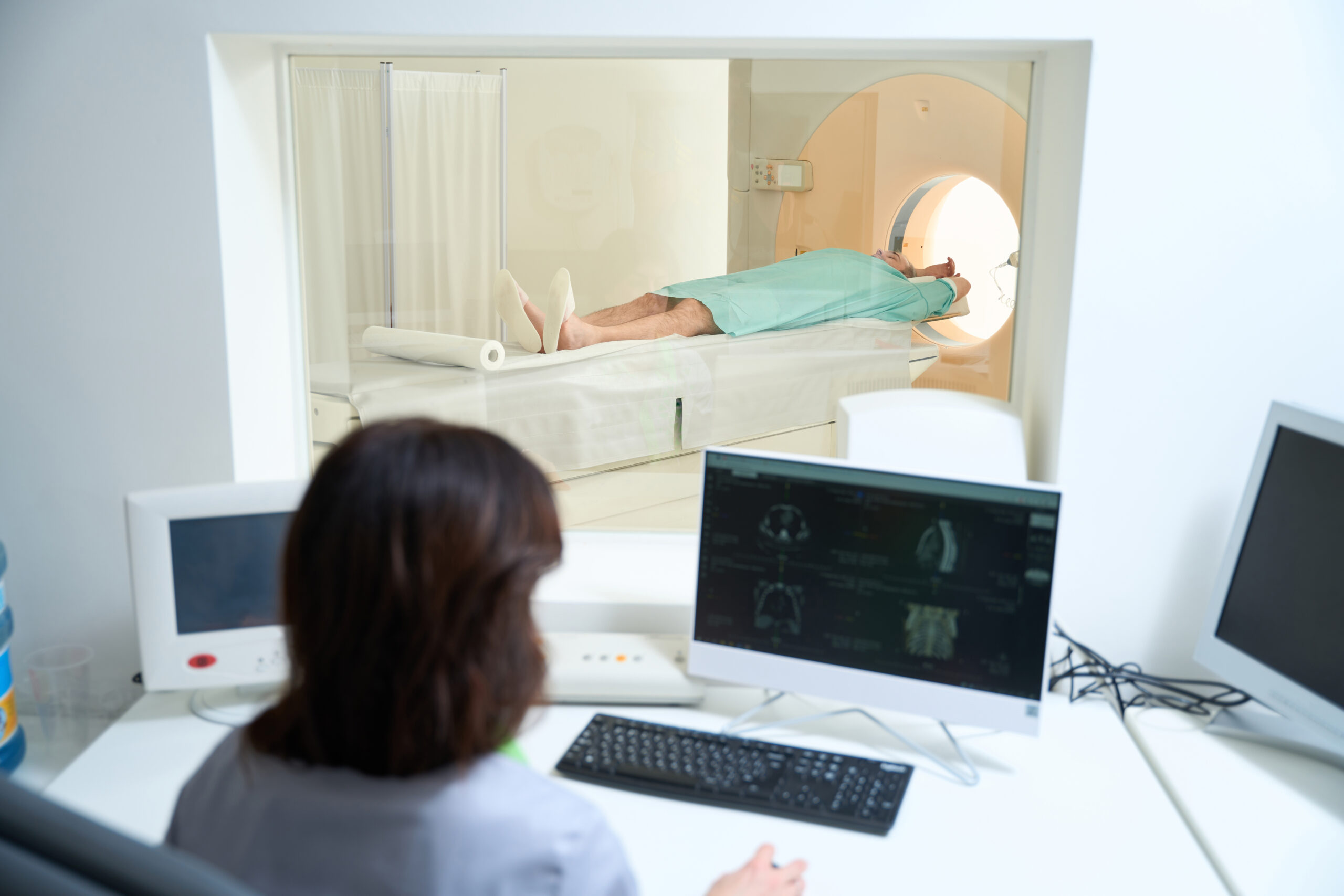
Unlike X-rays and CT scans, MRIs don’t use any radiation but rather magnetic fields and a sophisticated computer to take high-resolution pictures of your bones and soft tissues. A constant magnetic field and radio frequencies bounce off the fat and water molecules in your body. Radio waves are transmitted to a receiver in the machine which is translated into an image of the body.
“Often, problems are too subtle to see on an X-ray or may not be best seen on a CT image,” says Garner. “That’s where MRI comes in. They are especially useful for spotting sports injuries and musculoskeletal conditions. If your doctor sends you for an MRI, it may be to get a close look at tumors or cysts, get a closer look at your brain activity, evaluate certain types of heart issues, or examine certain diseases of internal organs.”
An MRI scanning procedure is painless, but it typically takes longer than X-rays and CT scans. You will lie on a flat, sliding bed that will pass into a tube-shaped MRI scanner. The magnet in the MRI machine produces repetitive thumping and tapping noises. You will be offered ear plugs during the scans. You will not feel any pain while undergoing an MRI, but the machine may be noisy.
Like a CT scan, an MRI scan may be done in a hospital or at an outpatient imaging center. Both CT scans and MRIs pose some risks when used. The risks are based on the type of imaging as well as how the imaging is performed.
The right imaging can lead to the right diagnosis, which is an essential part of effective treatment. It’s important to choose an imaging center that offers a full range of technologies, as well as experienced Radiologists and Technologists who are trained in specific imaging modalities (types), body areas, diseases and imaging techniques.
If your physician decides you need imaging, West Tennessee Healthcare is here to help. To make an Imaging Center appointment, click here.